
Health Facts

Back pain is often caused by obesity
Most people know that obesity contributes to the development of various diseases. However, did you know that obesity is a contributing factor to back pain? It is true. Being overweight or obese can significantly contribute to osteoporosis, osteoarthritis, rheumatoid arthritis, degenerative disc disease, spinal stenosis, and spondylolisthesis.
Procedure
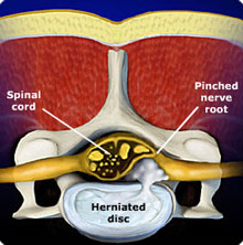
A disc ruptures when there is a tear in the outer lining (annulus) of the disc . When a tear in the annulus occurs, a fragment of disc material may protrude and pinch surrounding nerves. When a nerve is compressed it can cause symptoms such as extremity pain, numbness, weakness, electrical sensations, and bowel and bladder incontinence. If symptoms are not relieved with conservative treatments, a patient may be a candidate for surgical removal of the herniated disc fragment.Microendoscopic Discectomy differs from the open microdiscectomy. The incision using the microendoscopic technique is smaller (approximately 1cm), causing less trauma to the surrounding tissue. A smaller incision allows for decreased post-operative pain and a faster recovery. A patient is considered a potential candidate for a microendoscopic discectomy if he or she has a large herniated disc fragment extruded to the side of the spinal canal.
Microendoscopic Discectomy is performed by making a small incision in the patient's back and inserting a small endoscopic probe between the vertebrae and into the herniated disc space. A small camera is placed through the probe enabling the surgeon to view the operation on a TV screen in the operating room. Small surgical devices are placed through the probe to remove bone and herniated disc fragments.
The procedure usually takes about one hour; the patient is often able to return home on the same day. It is normal for a patient to experience postoperative pain, such as back pain, spasms, and lower extremity symptoms. These symptoms will usually improve as the nerve heals and inflammation of the nerve decreases. Patients are given pain medications during the healing process.
Latest news
Visit our media library for access to all of our news videos.
The Spine Institute is often in the news pioneering new treatments to help the reported 34 million Americans 18 years and older who suffer lower back pain, and another 9 million who suffer neck pain. Watch the news coverage here.